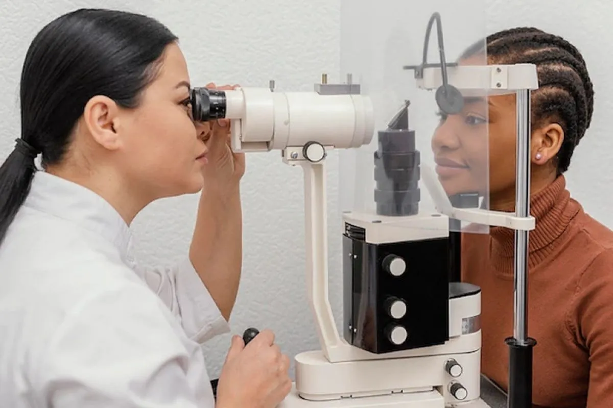
Advanced imaging techniques have transformed ophthalmology, enhancing the diagnosis and management of eye conditions. Optical coherence tomography (OCT), fundus photography, and confocal microscopy provide detailed eye anatomy and pathology visualizations. This article overviews these advancements, detailing each technique’s clinical applications. It aims to inform medical professionals and patients about innovations in eye care, from early detection of retinal diseases to precise corneal assessments. Explore the future of eye care and the invaluable insights these imaging techniques offer for accurate diagnoses and personalized treatment plans.
Importance Of Advanced Imaging In Diagnosing Eye Conditions
Advanced imaging in ophthalmology is vital for accurately diagnosing and managing eye diseases, significantly improving treatment outcomes through early detection. These techniques provide high-resolution images, allowing for the early identification of abnormalities in conditions like glaucoma, macular degeneration, and diabetic retinopathy.
They also enhance monitoring and treatment with quantitative data and visual evidence of changes, facilitating personalized care. For example, OCT scans detect subtle retinal changes, guiding therapy adjustments. Additionally, advanced imaging is an educational tool for medical professionals and students, enhancing their understanding of complex ocular pathologies.
Types Of Advanced Imaging Techniques Used In Ophthalmology
Ophthalmology employs various advanced imaging techniques, each with unique capabilities and applications. These techniques are categorized into structural, functional, and angiographic. Structural imaging focuses on the eye’s anatomy, utilizing methods like optical coherence tomography (OCT) for high-resolution cross-sectional images of the retina. Fundus photography captures the entire posterior segment, while confocal scanning laser ophthalmoscopy (CSLO) provides detailed pictures of retinal layers.
Angiographic techniques, including fluorescein and indocyanine green angiography, visualize retinal blood vessels to detect abnormalities like leaks or blockages. Additionally, ultrasonography is essential for examining ocular structures when direct visualization is hindered. Advanced imaging modalities such as MRI at Tellica Imaging further enhance diagnostic capabilities in ophthalmology. Understanding these techniques enables healthcare providers to select the most effective methods for diagnosing and managing eye conditions.
Optical Coherence Tomography (OCT) In Ophthalmology
Optical coherence tomography (OCT) is a crucial non-invasive imaging technique in ophthalmology that uses light waves to produce high-resolution, cross-sectional retina images. It aids in diagnosing and monitoring conditions like age-related macular degeneration (AMD), diabetic retinopathy, and glaucoma by detecting subtle changes that traditional methods might miss.
OCT measures retinal thickness and assesses the nerve fiber layer, enabling earlier interventions and personalized treatments. Recent advancements, including spectral-domain and swept-source OCT, enhance diagnostic capabilities with faster acquisition times and deeper choroid imaging, further improving the understanding and management of retinal diseases.
Fundus Photography And Its Applications In Ophthalmology
Fundus photography is an essential ophthalmology imaging technique that captures high-resolution images of the eye’s interior, including the retina, optic disc, and macula. It documents ocular findings, monitors disease progression, and aids in diagnoses.
Providing a permanent record allows for easy comparison over time, which is especially important for conditions like diabetes and hypertensive retinopathy. Regular imaging helps assess treatment effectiveness and adjust plans as needed.
Advancements like wide-field imaging and color fundus photography enhance its utility, enabling visualization of larger retinal areas and improving image detail and contrast, making it a vital tool for ophthalmologists.
Confocal Scanning Laser Ophthalmoscopy (CSLO) And Its Role In Diagnosing Eye Diseases
Confocal scanning laser ophthalmoscopy (CSLO) is a high-resolution retinal imaging technique that uses a laser to capture images at various depths. It is beneficial for assessing glaucoma by measuring nerve fiber thickness.
CSLO produces three-dimensional images that help diagnose retinal conditions like detachment, epiretinal membranes, and macular holes, revealing changes not seen with traditional methods. It can also be combined with autofluorescence and indocyanine green angiography for enhanced insights into retinal health, making it vital for managing complex retinal disorders.
Angiography Techniques For Assessing Retinal Blood Vessels
Angiography techniques are crucial for evaluating retinal blood vessels, primarily using fluorescein (FA) and indocyanine green angiography (ICG).
FA involves injecting fluorescein dye to image retinal vessels, aiding in diagnosing diabetic retinopathy. ICG uses indocyanine green dye to visualize choroidal vessels, helping assess conditions like choroidal neovascularization in age-related macular degeneration (AMD).
Both methods enhance the diagnosis and management of retinal diseases by providing real-time blood vessel visualization, leading to improved treatment strategies and outcomes.
Ultrasonography In Ophthalmology And Its Uses
Ultrasonography is a valuable ophthalmic imaging technique that uses high-frequency sound waves to visualize eye structures. It is beneficial for ophthalmologists in Troy, NY, when direct visualization is challenging, such as with cataracts or vitreous hemorrhage. This technique primarily assesses intraocular masses, measuring tumors’ size, shape, and location with B-scan ultrasonography to aid diagnosis and treatment planning. Additionally, it evaluates the anterior segment of the eye and assists with conditions like retinal detachment and foreign body localization. Advancements like high-frequency ultrasound enhance detail and resolution, solidifying ultrasonography as an essential tool for ophthalmologists.
Advancements In Imaging Technology For Ophthalmology
Ophthalmic imaging is advancing rapidly due to technological innovations and improved understanding of ocular diseases. Key developments include integrating artificial intelligence (AI) and machine learning, enhancing diagnostic accuracy, and early detection of eye conditions.
Wide-field imaging systems allow comprehensive evaluations of large retinal areas in a single image, which is crucial for assessing conditions like diabetic retinopathy.
Portable imaging devices increase access to advanced imaging in various settings, ensuring underserved communities receive quality eye care.
The continued evolution of imaging technology promises further advancements in personalized medicine and patient care, transforming the future of eye care.
Conclusion: The Future Of Advanced Imaging Techniques In Ophthalmology
In conclusion, advanced imaging techniques have transformed ophthalmology by enhancing the diagnosis and management of eye conditions. Techniques such as optical coherence tomography, fundus photography, confocal scanning laser ophthalmoscopy, and angiography provide crucial insights and support personalized treatments.
Future advancements, including artificial intelligence and portable imaging devices, promise to improve patient outcomes through timely interventions. Staying informed about these innovations is essential for medical professionals and patients alike, as they will optimize eye care and usher in a new era of enhanced vision and eye health.